Ultrasound
Ultrasound is a safe, non-invasive imaging exam that uses sound waves to create real-time images of the body’s internal structures. It is widely used to evaluate organs and soft tissues without the use of radiation.
Ultrasound is commonly used to examine:
- Liver, gallbladder, and kidneys
- Thyroid
- Uterus and ovaries
- Testicles
- Arteries and veins
- Pregnancy and fetal development
Preparation
Preparation depends on the type of exam:
- Abdominal ultrasound: Fasting for 4–6 hours is typically required
- Pelvic ultrasound: You may be asked to arrive with a full bladder to improve image quality
Your care team will provide specific instructions before your appointment.
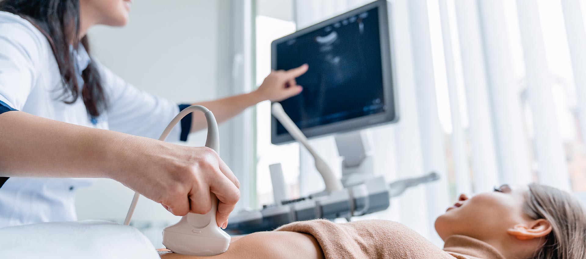
What to Expect
During the exam, a technologist will gently place a handheld probe on the area being imaged. Ultrasound exams are painless and typically take 15–30 minutes to complete.
For certain pelvic exams, a transvaginal ultrasound may be recommended to obtain more detailed images of the uterus and ovaries. This is a commonly performed procedure and is generally well tolerated. The additional detail it provides is often essential for accurate diagnosis.
Safe, Real-Time Imaging
Ultrasound offers the advantage of real-time visualization, allowing physicians to assess structure and function simultaneously—supporting timely, accurate clinical decisions.
For more information about CT scans and other imaging procedures, radiologyinfo.org